
Lower Blepharoplasty with Fat Transposition Canthopexy
Before & after upper lower blepharoplasty with fat pad transposition and canthopexy in a 44-year-old. 3-week results from board-certified surgeon in Istanbul
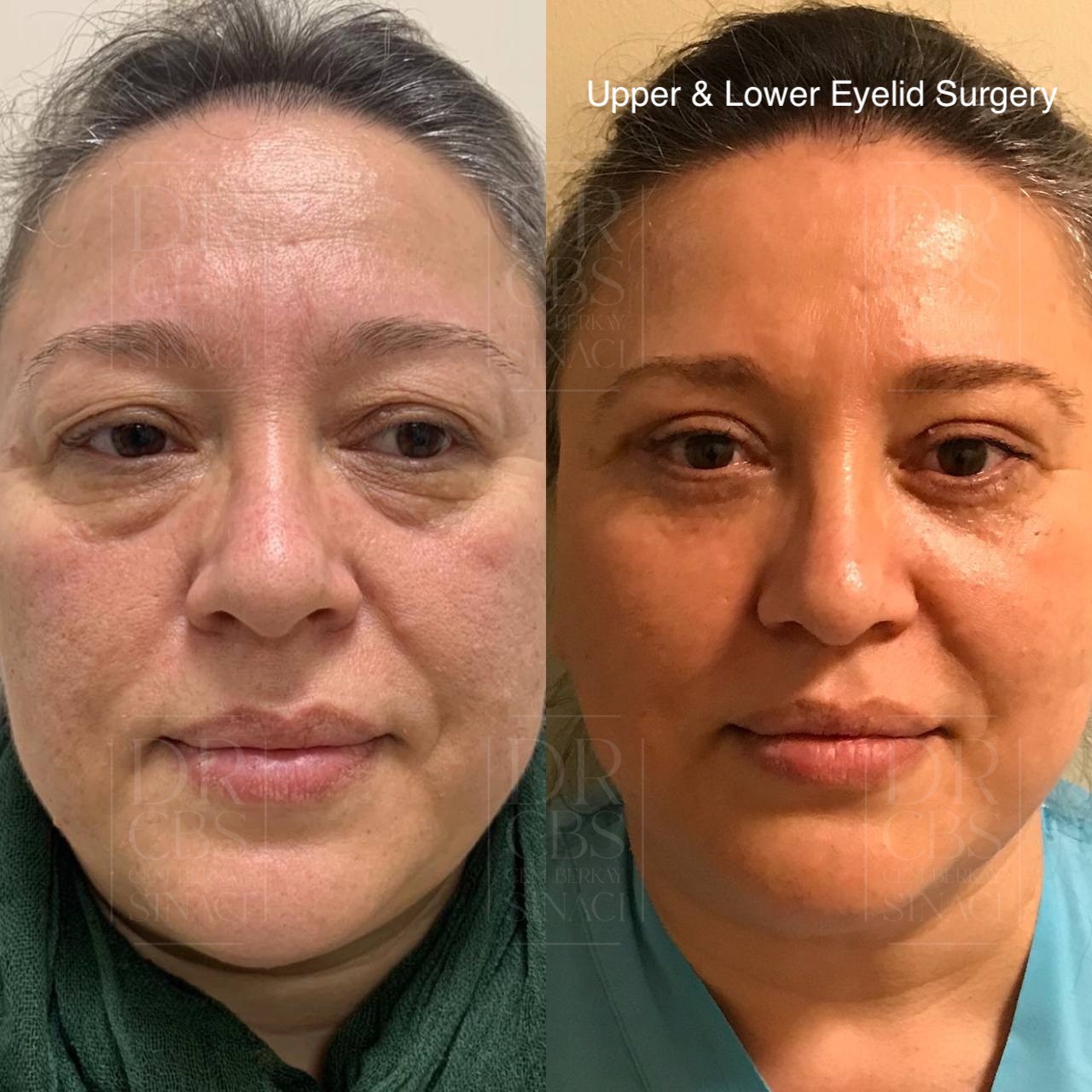
Patient Overview
Patient: Huda
Age: 44
Gender: Female
Procedures: Upper blepharoplasty, lower blepharoplasty with fat pad transposition, canthopexy
After photos taken at: 3 weeks post-op
Location: Istanbul, Turkey
The Difference Between Removing Fat and Repositioning It
For decades, lower blepharoplasty meant one thing: cutting away the fat bags beneath the eyes. The approach was effective at eliminating puffiness, but it often created a new problem — a hollow, skeletonised appearance that could make the under-eye area look older rather than younger. Modern lower eyelid surgery has evolved beyond simple removal. In Huda's case, Dr. Cem Berkay Sinaci, a European board-certified plastic surgeon (FEBOPRAS) and member of ISAPS and ASPS, used fat pad transposition — a technique that keeps the patient's own fat attached to its blood supply and repositions it downward to fill the tear trough depression, effectively solving two problems with the same tissue.
What Is Fat Pad Transposition?
The lower eyelid contains three distinct fat compartments — medial, central, and lateral — separated by thin connective tissue walls called septae. When these fat pads herniate forward due to ageing or genetic predisposition, they create the visible bags and puffiness beneath the eyes. Simultaneously, the area just below these bags — the tear trough — often appears hollow and shadowed, creating a tired, aged look.
Fat pad transposition addresses both concerns in a single manoeuvre. Rather than excising the herniated fat, the surgeon releases it from its compartment while maintaining its vascular pedicle, then redirects it inferiorly to drape over the orbital rim and fill the tear trough hollow. The fat is secured in its new position with a small external fixation suture placed on the cheek skin, which holds everything precisely in place during the initial healing period. This external stitch is removed one week later in a simple clinic visit.
Huda, a 44-year-old female patient, had both prominent lower eyelid fat bags and noticeable tear trough depressions — making her an ideal candidate for this redistribution approach rather than simple excision.
Why Canthopexy Was Performed Alongside
Any time the lower eyelid is opened surgically, there is a temporary period of reduced lid tone during healing. In patients who have even mild pre-existing laxity of the lower lid, this can result in the lid pulling downward — a complication known as lower lid retraction or ectropion. Canthopexy prevents this by tightening the lateral canthal tendon at the outer corner of the eye, reinforcing the lower lid's suspension before the surgical swelling and healing process begins.
In Huda's case, canthopexy served a dual purpose. Beyond its protective function, it helped create a subtle positive canthal tilt — where the outer corner of the eye sits slightly higher than the inner corner. This is a hallmark of a youthful eye shape and one of the details that separates a technically competent blepharoplasty from an aesthetically refined one. Having trained in advanced facial rejuvenation techniques through fellowship in Brazil and cadaver dissection in Bangkok, Dr. Sinaci considers canthopexy an integral component of subciliary lower blepharoplasty rather than an optional addition.
The Upper Blepharoplasty Component
Huda's upper eyelids showed the typical changes of the mid-forties — excess skin folding over the natural crease, creating a hooded appearance that narrowed the visible lid platform. This was corrected through a standard upper blepharoplasty incision placed within the eyelid crease. The redundant skin and a small strip of underlying muscle were excised, restoring a clean, defined crease that is concealed when the eyes are open.
Performing both upper and lower blepharoplasty in a single session offers the advantage of a unified recovery period and a balanced result. Correcting only one set of lids can sometimes make the untreated pair appear comparatively worse, so addressing both simultaneously ensures the entire periorbital region rejuvenates together.
Non-Dissolvable Sutures in Eyelid Surgery
For all incision closures — both upper lids and the subciliary lower lid incisions — Dr. Sinaci used non-dissolvable suture material. Eyelid skin is extraordinarily thin and reacts visibly to any inflammation within it. Dissolvable sutures break down through an enzymatic or hydrolytic reaction that triggers an inflammatory response in the surrounding tissue, and in skin this thin, that reaction can translate into wider, more noticeable scars. Non-dissolvable sutures sit inertly in the tissue, cause essentially no inflammatory reaction, and are removed between four and six days after surgery. The trade-off of one brief clinic visit produces meaningfully better scar quality.
Understanding the Three-Week Recovery Stage
Huda's before and after photographs at three weeks capture an honest mid-recovery moment. By this stage, the most visible bruising has resolved and the major swelling has subsided, but the tissues are still actively healing. There may be mild residual puffiness, particularly in the lower lids where the fat transposition and canthopexy create slightly more tissue manipulation than a simple upper blepharoplasty alone. The external cheek fixation suture for the transposed fat was removed at one week, and by three weeks the small mark it leaves has typically faded or is fading.
What is already clearly visible at this stage is the improvement in overall eye contour. The upper lid hooding has been corrected, revealing a defined crease. The lower lid bags are absent, and — critically — the tear trough area that was previously hollow now has a smooth, filled transition from lower lid to cheek. This seamless lid-cheek junction is the signature advantage of fat transposition over simple fat removal.
The Value of Using Your Own Tissue
Fat pad transposition offers something that no injectable filler can replicate with the same permanence or naturalness. The repositioned fat maintains its own blood supply, meaning it behaves as living tissue in its new location — it does not reabsorb, migrate, or require repeat treatments. Dermal fillers placed in the tear trough can produce excellent short-term results, but they are temporary, carry the risk of vascular complications in this highly vascular region, and can accumulate over repeated sessions to create a puffy or unnatural appearance. By using the tissue that is already there — simply in the wrong position — fat transposition provides a one-time, permanent correction with a completely natural feel and appearance.
Choosing Combined Blepharoplasty in Istanbul
For patients researching eye lift surgery in Turkey, Huda's case demonstrates what a comprehensive approach to periorbital rejuvenation looks like. This was not a single-technique procedure — it involved four distinct surgical steps across upper and lower lids, each addressing a specific anatomical concern. The combination of upper blepharoplasty, lower lid fat transposition, skin removal, and canthopexy required planning that accounts for how each element interacts with the others. At three weeks, Huda's result already shows the refreshed, natural appearance that will continue to refine over the coming months as the final healing matures.