
Otoplasty Ear Pinning Before and After at 2 Weeks
Before and after otoplasty ear pinning and reshaping at two weeks post-surgery. Dr. Cem Berkay Sinaci explains ear surgery recovery in Istanbul.
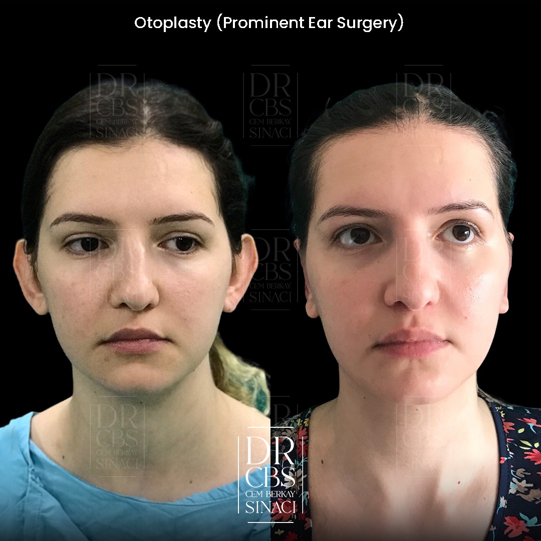
Patient Overview
Patient: Nili
Age: 21 years old
Gender: Female
Procedures: Otoplasty (ear pinning and reshaping)
After photos taken at: 2 weeks post-surgery
Location: Istanbul, Turkey
Living with Prominent Ears: Why Patients Seek Otoplasty
Prominent ears — ears that project further from the head than the aesthetic norm — are one of the earliest physical features a person becomes conscious of. Unlike breast size or nasal shape, which typically become concerns during adolescence or adulthood, ear prominence is noticed in childhood, often by peers before the individual themselves. The psychological impact can begin remarkably early and persist for decades, influencing hairstyle choices, social behaviour, and self-consciousness in ways that outsiders may never suspect.
Nili, a twenty-one-year-old patient of Dr. Cem Berkay Sinaci, made the decision to address her prominent ears at an age when she could choose the procedure for herself with full understanding of what it involves and what it achieves. While otoplasty can be performed on children as young as five or six — once the ear cartilage has reached near-adult size — many patients prefer to wait until adulthood, when the decision is entirely their own.
Dr. Sinaci, a European board-certified plastic surgeon (FEBOPRAS) and active member of ISAPS and ASPS, performs otoplasty for both adults and children, tailoring the technique to the specific anatomical features that contribute to each patient's ear prominence. The goal is never to create ears that look "operated" — pinned flat against the head in an unnatural position — but to produce ears that fall within the normal range of projection, appearing as though they simply grew this way.
The Anatomy Behind Prominent Ears
Ear prominence is not a single condition but a result of one or more anatomical variations that can occur independently or in combination. Understanding which structures are contributing to the projection is essential for planning a correction that addresses the root cause rather than simply forcing the ear backward.
The antihelical fold is the most common culprit. This Y-shaped cartilage ridge runs vertically through the central portion of the ear, creating the natural contour that angles the upper ear toward the head. When the antihelical fold is underdeveloped or absent, the upper ear lacks this natural bend and projects outward, creating the appearance of ears that stick out from the head.
The conchal bowl — the deep, cup-shaped cartilage directly surrounding the ear canal — can also contribute to prominence when it is excessively deep or large. Even with a normal antihelical fold, an oversized concha pushes the entire ear away from the skull, producing projection that originates from the base of the ear rather than the upper portion.
In some patients, both factors are present. Nili's preoperative assessment identified the specific anatomical contributors to her ear prominence, allowing Dr. Sinaci to design a surgical plan that addressed each element individually while producing a naturally contoured result.
What Otoplasty Surgery Involves
Otoplasty is performed through an incision placed behind the ear — in the natural crease where the ear meets the scalp. This location ensures that the surgical scar is virtually invisible, hidden in a fold that is never exposed during normal social interaction.
Through this incision, the ear cartilage is accessed and reshaped using a combination of techniques selected for the specific anatomical correction required. When the antihelical fold needs to be created or enhanced, the cartilage is scored, folded, and secured with permanent sutures that hold the new contour in place while the cartilage heals into its reshaped position. When conchal reduction is necessary, a carefully calculated segment of conchal cartilage is removed and the remaining edges are sutured together, reducing the depth of the bowl and bringing the ear closer to the head.
The artistry of otoplasty lies in calibration. The surgeon must determine precisely how much correction to apply — enough to bring the ears into a natural range of projection, but not so much that they appear pinned or flattened. Overcorrection is as aesthetically displeasing as the original prominence, and far more difficult to revise. Dr. Sinaci's approach to this calibration, refined through cadaver training in Bangkok and years of clinical experience, prioritises a result that looks uncontrived — ears that appear naturally positioned rather than surgically altered.
The Two-Week Result: Early but Telling
Nili's two-week photographs show ears that have already achieved their corrected position. Unlike procedures where the final result emerges gradually over months, otoplasty produces its fundamental shape change immediately — the cartilage is reshaped and sutured into its new configuration during surgery, and this new configuration is present from the moment the procedure is complete.
What changes during the healing period is not the ear position but the tissue quality surrounding it. At two weeks, residual swelling is still present in the ear cartilage and the surrounding skin. The ears may appear slightly thicker and less defined in their contour than they will at the final result. The skin colour may still show traces of bruising, though this varies considerably between patients. Sensation in the ears may be altered — numbness, tingling, or hypersensitivity are all normal at this stage and resolve progressively over weeks to months.
Despite these expected healing phenomena, the corrected ear projection is clearly visible at two weeks. The ears sit closer to the head, the antihelical fold — if this was part of the correction — creates a natural contour that breaks up the smooth, projected surface of the uncorrected ear, and the overall profile shows ears that fall within normal aesthetic proportions.
The Headband Protocol: Why It Matters
Post-operative protection is critical to the long-term success of otoplasty, and the headband protocol that Dr. Sinaci prescribes is a non-negotiable element of the recovery plan. The reshaped cartilage has been sutured into its new position, but the tissue requires time to heal and stabilise in this configuration. During this period, any force that bends the ear forward — such as the pressure of a pillow during sleep — can stress the sutures and potentially compromise the correction.
Dr. Sinaci recommends two weeks of continuous headband use, worn day and night. The headband holds the ears gently against the head, protecting the healing cartilage from accidental trauma during daily activities and from the unconscious pressure of sleeping positions. During these first two weeks, when the surgical repair is most vulnerable, consistent headband wear provides the mechanical protection that allows the internal sutures to do their job without external interference.
After the initial two-week period, the headband transitions to nighttime use only, worn during sleep for an additional month. This extended night protection addresses a specific risk: during sleep, patients turn unconsciously, and the ear can fold forward against the pillow with sustained pressure that the sleeping patient is unaware of. A month of nighttime headband use protects the ears during this vulnerable period until the cartilage has healed firmly enough to resist bending forces on its own.
This six-week total headband protocol — two weeks continuous, one month nighttime — may seem demanding, but it represents a small investment of discipline that protects the surgical result for a lifetime. Patients who comply fully with the headband protocol give their cartilage the best possible conditions to heal in its corrected position permanently.
Otoplasty for Adults Versus Children
While otoplasty is frequently associated with childhood correction — and indeed many parents seek the procedure for children who are experiencing social difficulty due to prominent ears — adult otoplasty is equally common and produces excellent results. There is, however, a biological distinction worth understanding. Children's ear cartilage is softer and more pliable, which means it accepts reshaping more readily and, crucially, has less cartilage memory. Once reshaped and sutured into the corrected position, the younger cartilage is more likely to remain there permanently, resulting in lower recurrence rates.
Adult cartilage, by contrast, is firmer and carries stronger structural memory — an inherent tendency to return toward its original shape. This cartilage memory means that recurrence rates in adult otoplasty are slightly higher than in children. The recurrence remains rare, but it is the primary reason why the headband protocol is so important in adult patients: the external support during the healing period counteracts the cartilage's tendency to spring back while the internal sutures and scar tissue formation lock the new shape into place.
This biological reality is also why Dr. Sinaci supports early otoplasty for children whose prominent ears are causing psychological distress. Correcting the ears when the cartilage is most receptive to permanent reshaping — typically around age five to six, when the ear has reached approximately ninety percent of its adult size — offers the best long-term structural outcome while simultaneously sparing the child years of self-consciousness.
Nili's decision to undergo otoplasty at twenty-one reflects a pattern Dr. Sinaci sees regularly: adults who endured years of self-consciousness about their ears during childhood and adolescence, adapting their hairstyles and behaviour to conceal the prominence, and who finally choose correction when they have the autonomy and resources to do so. Despite the marginally higher recurrence risk compared to childhood surgery, adult otoplasty remains highly successful, and the emotional relief these patients experience often surpasses what might be expected for what is, surgically, a relatively modest procedure.
Scarring and Long-Term Appearance
The otoplasty incision, placed in the posterior auricular crease, heals with a scar that is remarkably well concealed. This anatomical location benefits from excellent blood supply, consistent moisture levels, and freedom from the tension forces that can widen scars in other body regions. By six months, the posterior auricular scar is typically a thin, pale line that is invisible unless the ear is deliberately folded forward for inspection.
The ear contour itself continues to refine subtly over the first three to six months as residual cartilage swelling resolves and the skin re-drapes over the reshaped framework. The final result — which Nili will appreciate fully by six months — shows ears with natural folds, proportional projection, and symmetry that appears entirely innate.
Otoplasty in Istanbul
Nili's two-week before and after photographs demonstrate the immediate and visible impact of otoplasty on ear prominence. For adults and children considering ear reshaping surgery in Istanbul, her case illustrates that the correction is established at surgery and visible from the earliest post-operative days, with continued refinement as healing progresses. The headband protocol — two weeks continuous wear followed by one month of nighttime use — is a simple but essential element that protects the result and ensures the reshaped cartilage heals in its corrected position permanently.