
Lower Blepharoplasty for Under-Eye Bags at 31
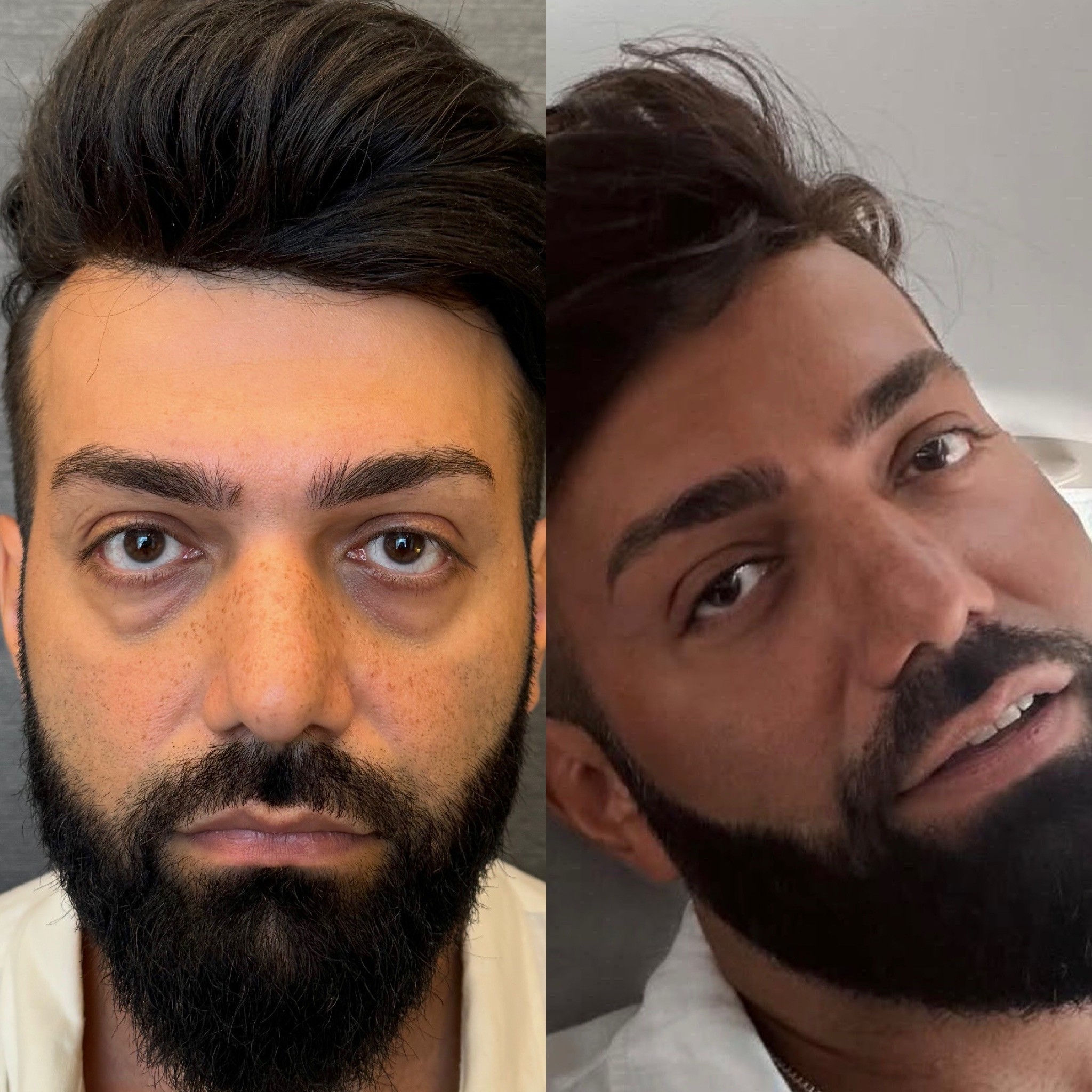
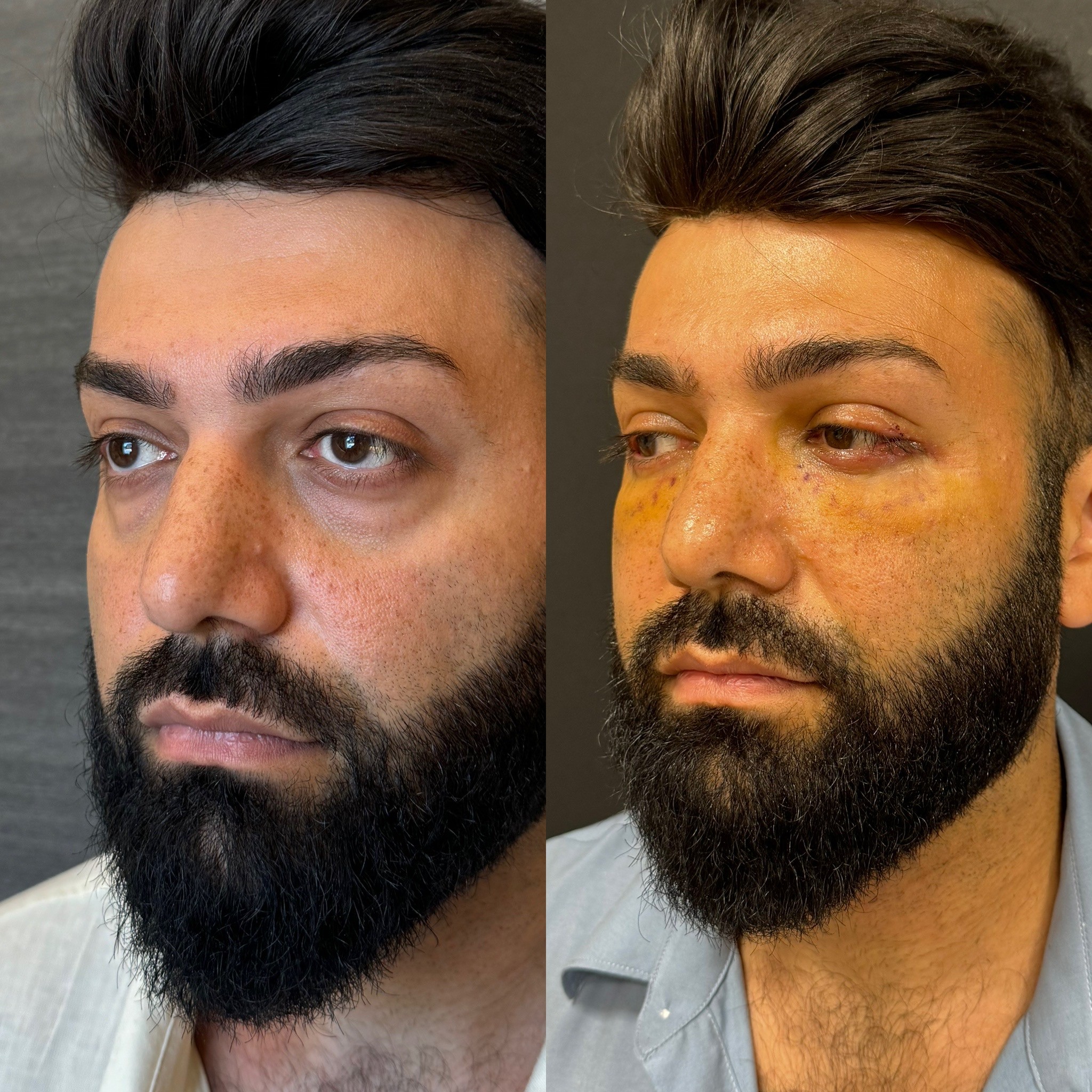
Before & after lower blepharoplasty for under-eye bags and scleral show correction at 6 months and and five days. Dr. CBS lower eyelid surgery in Istanbul.

Patient Overview
Patient: Vladimir
Age: 31 years old
Gender: Male
Procedures: Lower blepharoplasty (subciliary approach) — under-eye bag removal and scleral show correction
After photos taken at: 5 days post-surgery
Location: Istanbul, Turkey
Under-Eye Bags at Thirty-One: When It Is Not About Sleep
The advice is always the same — sleep more, drink water, reduce stress, try a cold compress. Patients with under-eye bags hear these suggestions constantly from well-meaning friends who assume the puffiness is a lifestyle problem. For some, it is. For Vladimir, a thirty-one-year-old male patient of Dr. Cem Berkay Sinaci, it was not. His under-eye bags were structural — herniated orbital fat pushing forward through a weakened orbital septum, creating permanent convexities beneath both eyes that no amount of rest, hydration, or eye cream would ever reduce.
Dr. Sinaci, a European board-certified plastic surgeon (FEBOPRAS) and member of ISAPS and ASPS, confirmed during consultation what Vladimir already suspected: the bags were anatomical, present regardless of sleep quality or lifestyle, and correctable only through lower blepharoplasty. At thirty-one, Vladimir is younger than the typical lower eyelid surgery patient — but under-eye fat herniation is a genetic trait that can manifest well before middle age, and waiting for it to worsen serves no clinical purpose.
What Causes Under-Eye Bags
The lower eyelid sits over a layer of orbital fat that cushions and protects the eyeball within its bony socket. This fat is held in position by a thin membrane called the orbital septum — a fascial barrier that keeps the fat pads contained behind the eyelid surface.
In patients with under-eye bags, the orbital septum has weakened or thinned, allowing the fat pads behind it to push forward. The fat itself has not increased in volume — it has simply migrated anteriorly through a barrier that no longer contains it. The result is the characteristic bulging beneath the eye that creates shadows, disrupts the smooth lower eyelid contour, and produces the tired, aged appearance that patients describe.
This weakening can be age-related — the septum thins gradually over decades — or genetic, where the septum is constitutionally thinner from youth. Vladimir's presentation at thirty-one places him firmly in the genetic category. His under-eye bags were not a product of ageing but of inherited septal anatomy that allowed early fat herniation.
The Subciliary Approach: Invisible Access
Dr. Sinaci performed Vladimir's lower blepharoplasty through a subciliary incision — a cut placed directly beneath the lower eyelash line. This approach provides full access to the lower eyelid fat compartments, the orbital septum, and the deeper structural layers that must be addressed during comprehensive lower blepharoplasty.
The subciliary incision follows the natural curve of the lower lash margin, sitting one to two millimetres below the eyelash roots. This placement exploits the visual camouflage that the lash line provides — the incision hides in the shadow cast by the lashes themselves, and once healed, the scar blends into the delicate skin texture of the lower eyelid margin where it becomes essentially undetectable.
Alternative approaches exist — the transconjunctival approach places the incision inside the lower eyelid, leaving no external scar at all. But the subciliary approach offers broader access for cases that require not only fat removal or repositioning but also skin excision, muscle tightening, or structural reinforcement of the lower eyelid. For Vladimir's combination of fat herniation and scleral show correction, the subciliary approach provided the comprehensive access necessary to address both concerns through a single incision.
Scleral Show Correction: The Structural Component
Vladimir's surgical plan included correction of scleral show — a condition where a visible strip of white sclera appears between the lower iris and the lower eyelid margin. In a normally positioned lower eyelid, the lid margin touches or slightly overlaps the lower edge of the iris. When the lower eyelid sits lower than this ideal position, the exposed sclera creates a rounded, startled appearance and can contribute to the tired look that under-eye bags already produce.
Scleral show correction through lower blepharoplasty involves tightening and repositioning the lower eyelid structures to elevate the lid margin to its ideal position against the globe. This may include lateral canthal tightening — reinforcing the outer corner attachment of the lower eyelid to the orbital rim — and careful management of the skin-muscle layers to support the eyelid in its corrected position.
This structural component adds complexity to the procedure beyond simple fat removal. It requires precise understanding of lower eyelid mechanics — the balance between the forces that hold the eyelid against the eye and the forces that pull it downward. Overcorrection risks ectropion, where the eyelid turns outward, exposing the conjunctiva. Undercorrection leaves residual scleral show that fails to meet the surgical objective.
Dr. Sinaci's approach to lower eyelid positioning, refined through his fellowship with the internationally renowned plastic surgeon Raul Gonzalez in Brazil and cadaver dissection training in Bangkok, calibrates the structural tightening to produce a natural lid position — snug against the globe without excessive tension that could compromise eyelid function.
Day Five: Honest Early Recovery
Vladimir's five-day photographs show the lower blepharoplasty in its acute post-operative state. Interpreting these images requires understanding that day five represents the body's most active healing response to lower eyelid surgery.
Swelling is the dominant feature. The lower eyelids carry significant oedema that temporarily distorts the contour the surgery created. The tissue beneath the eyes appears puffy — paradoxically similar to the bags that were just removed — but this puffiness is inflammatory fluid, not herniated fat. It is temporary, and it will resolve predictably over the next two to three weeks.
The subciliary incision is visible at five days as a fine line beneath the lash margin. Sutures may still be present or recently removed. The incision line will progressively fade over the coming weeks and months, eventually becoming indistinguishable from the natural lower lash line texture.
Bruising at five days may extend to the lower eyelid, upper cheek, and sometimes the medial corner of the eye. The extent varies between patients, but the resolution follows a predictable timeline — colour changes from dark to yellow-green over seven to fourteen days before clearing completely.
The scleral show correction is not fully assessable at day five due to the swelling that temporarily affects lower eyelid position. The true lid position will become apparent as the oedema resolves, typically stabilising by four to six weeks.
Why Five-Day Documentation Matters
Post-operative photography at day five serves a specific educational purpose for lower blepharoplasty. This procedure generates more early recovery anxiety than upper blepharoplasty because the swelling pattern can temporarily mimic the very problem that was corrected. Patients who see puffy lower eyelids at day five — without understanding that this is inflammatory oedema, not recurrent bags — can experience significant distress.
Vladimir's five-day images provide future patients with an honest reference for what this stage looks like. The swelling is real, visible, and expected. It is not the result. It is the healing process that precedes the result. The distinction is critical, and having photographic evidence of another patient at this same stage normalises the experience.
The Weeks Ahead
Vladimir's lower blepharoplasty result will emerge progressively over the next four to eight weeks. The swelling resolves in waves — the most dramatic reduction occurs between days seven and fourteen, with continued refinement through week six. The under-eye contour that the surgery created — smooth, free of herniated fat bulges, with a natural transition from eyelid to cheek — becomes increasingly visible as each layer of oedema clears.
The scleral show correction stabilises by six weeks as the lower eyelid settles into its final supported position against the globe. The subciliary scar matures over three to six months, fading from its early pink appearance to a pale line hidden beneath the lashes.
By three months, Vladimir will see the definitive result — smooth lower eyelids, corrected lid position, and no visible scar — a face free of the genetic under-eye bags that no lifestyle change could address and that only surgical correction could resolve.
Lower Blepharoplasty in Istanbul
Vladimir's five-day before and after documents the honest early reality of subciliary lower blepharoplasty — a procedure that corrects under-eye bags and scleral show through an incision that heals to near-invisibility beneath the lash line. For patients researching lower eyelid surgery and under-eye bag removal in Istanbul, his case demonstrates that structural fat herniation is a genetic condition treatable at any age, and that the brief post-operative swelling at day five bears no resemblance to the smooth, refreshed lower eyelid contour that emerges in the weeks that follow.
Under-Eye Bags at Thirty-One: When It Is Not About Sleep
The advice is always the same — sleep more, drink water, reduce stress, try a cold compress. Patients with under-eye bags hear these suggestions constantly from well-meaning friends who assume the puffiness is a lifestyle problem. For some, it is. For Vladimir, a thirty-one-year-old male patient of Dr. Cem Berkay Sinaci, it was not. His under-eye bags were structural — herniated orbital fat pushing forward through a weakened orbital septum, creating permanent convexities beneath both eyes that no amount of rest, hydration, or eye cream would ever reduce.
Dr. Sinaci, a European board-certified plastic surgeon (FEBOPRAS) and member of ISAPS and ASPS, confirmed during consultation what Vladimir already suspected: the bags were anatomical, present regardless of sleep quality or lifestyle, and correctable only through lower blepharoplasty. At thirty-one, Vladimir is younger than the typical lower eyelid surgery patient — but under-eye fat herniation is a genetic trait that can manifest well before middle age, and waiting for it to worsen serves no clinical purpose.
What Causes Under-Eye Bags
The lower eyelid sits over a layer of orbital fat that cushions and protects the eyeball within its bony socket. This fat is held in position by a thin membrane called the orbital septum — a fascial barrier that keeps the fat pads contained behind the eyelid surface.
In patients with under-eye bags, the orbital septum has weakened or thinned, allowing the fat pads behind it to push forward. The fat itself has not increased in volume — it has simply migrated anteriorly through a barrier that no longer contains it. The result is the characteristic bulging beneath the eye that creates shadows, disrupts the smooth lower eyelid contour, and produces the tired, aged appearance that patients describe.
This weakening can be age-related — the septum thins gradually over decades — or genetic, where the septum is constitutionally thinner from youth. Vladimir's presentation at thirty-one places him firmly in the genetic category. His under-eye bags were not a product of ageing but of inherited septal anatomy that allowed early fat herniation.
The Subciliary Approach: Invisible Access
Dr. Sinaci performed Vladimir's lower blepharoplasty through a subciliary incision — a cut placed directly beneath the lower eyelash line. This approach provides full access to the lower eyelid fat compartments, the orbital septum, and the deeper structural layers that must be addressed during comprehensive lower blepharoplasty.
The subciliary incision follows the natural curve of the lower lash margin, sitting one to two millimetres below the eyelash roots. This placement exploits the visual camouflage that the lash line provides — the incision hides in the shadow cast by the lashes themselves, and once healed, the scar blends into the delicate skin texture of the lower eyelid margin where it becomes essentially undetectable.
Alternative approaches exist — the transconjunctival approach places the incision inside the lower eyelid, leaving no external scar at all. But the subciliary approach offers broader access for cases that require not only fat removal or repositioning but also skin excision, muscle tightening, or structural reinforcement of the lower eyelid. For Vladimir's combination of fat herniation and scleral show correction, the subciliary approach provided the comprehensive access necessary to address both concerns through a single incision.
Scleral Show Correction: The Structural Component
Vladimir's surgical plan included correction of scleral show — a condition where a visible strip of white sclera appears between the lower iris and the lower eyelid margin. In a normally positioned lower eyelid, the lid margin touches or slightly overlaps the lower edge of the iris. When the lower eyelid sits lower than this ideal position, the exposed sclera creates a rounded, startled appearance and can contribute to the tired look that under-eye bags already produce.
Scleral show correction through lower blepharoplasty involves tightening and repositioning the lower eyelid structures to elevate the lid margin to its ideal position against the globe. This may include lateral canthal tightening — reinforcing the outer corner attachment of the lower eyelid to the orbital rim — and careful management of the skin-muscle layers to support the eyelid in its corrected position.
This structural component adds complexity to the procedure beyond simple fat removal. It requires precise understanding of lower eyelid mechanics — the balance between the forces that hold the eyelid against the eye and the forces that pull it downward. Overcorrection risks ectropion, where the eyelid turns outward, exposing the conjunctiva. Undercorrection leaves residual scleral show that fails to meet the surgical objective.
Dr. Sinaci's approach to lower eyelid positioning, refined through his fellowship with the internationally renowned plastic surgeon Raul Gonzalez in Brazil and cadaver dissection training in Bangkok, calibrates the structural tightening to produce a natural lid position — snug against the globe without excessive tension that could compromise eyelid function.
Day Five: Honest Early Recovery
Vladimir's five-day photographs show the lower blepharoplasty in its acute post-operative state. Interpreting these images requires understanding that day five represents the body's most active healing response to lower eyelid surgery.
Swelling is the dominant feature. The lower eyelids carry significant oedema that temporarily distorts the contour the surgery created. The tissue beneath the eyes appears puffy — paradoxically similar to the bags that were just removed — but this puffiness is inflammatory fluid, not herniated fat. It is temporary, and it will resolve predictably over the next two to three weeks.
The subciliary incision is visible at five days as a fine line beneath the lash margin. Sutures may still be present or recently removed. The incision line will progressively fade over the coming weeks and months, eventually becoming indistinguishable from the natural lower lash line texture.
Bruising at five days may extend to the lower eyelid, upper cheek, and sometimes the medial corner of the eye. The extent varies between patients, but the resolution follows a predictable timeline — colour changes from dark to yellow-green over seven to fourteen days before clearing completely.
The scleral show correction is not fully assessable at day five due to the swelling that temporarily affects lower eyelid position. The true lid position will become apparent as the oedema resolves, typically stabilising by four to six weeks.
Why Five-Day Documentation Matters
Post-operative photography at day five serves a specific educational purpose for lower blepharoplasty. This procedure generates more early recovery anxiety than upper blepharoplasty because the swelling pattern can temporarily mimic the very problem that was corrected. Patients who see puffy lower eyelids at day five — without understanding that this is inflammatory oedema, not recurrent bags — can experience significant distress.
Vladimir's five-day images provide future patients with an honest reference for what this stage looks like. The swelling is real, visible, and expected. It is not the result. It is the healing process that precedes the result. The distinction is critical, and having photographic evidence of another patient at this same stage normalises the experience.
The Weeks Ahead
Vladimir's lower blepharoplasty result will emerge progressively over the next four to eight weeks. The swelling resolves in waves — the most dramatic reduction occurs between days seven and fourteen, with continued refinement through week six. The under-eye contour that the surgery created — smooth, free of herniated fat bulges, with a natural transition from eyelid to cheek — becomes increasingly visible as each layer of oedema clears.
The scleral show correction stabilises by six weeks as the lower eyelid settles into its final supported position against the globe. The subciliary scar matures over three to six months, fading from its early pink appearance to a pale line hidden beneath the lashes.
By three months, Vladimir will see the definitive result — smooth lower eyelids, corrected lid position, and no visible scar — a face free of the genetic under-eye bags that no lifestyle change could address and that only surgical correction could resolve.
Lower Blepharoplasty in Istanbul
Vladimir's five-day before and after documents the honest early reality of subciliary lower blepharoplasty — a procedure that corrects under-eye bags and scleral show through an incision that heals to near-invisibility beneath the lash line. For patients researching lower eyelid surgery and under-eye bag removal in Istanbul, his case demonstrates that structural fat herniation is a genetic condition treatable at any age, and that the brief post-operative swelling at day five bears no resemblance to the smooth, refreshed lower eyelid contour that emerges in the weeks that follow.