
Waterfall Deformity & Full Muscle Coverage Fix | Day 5
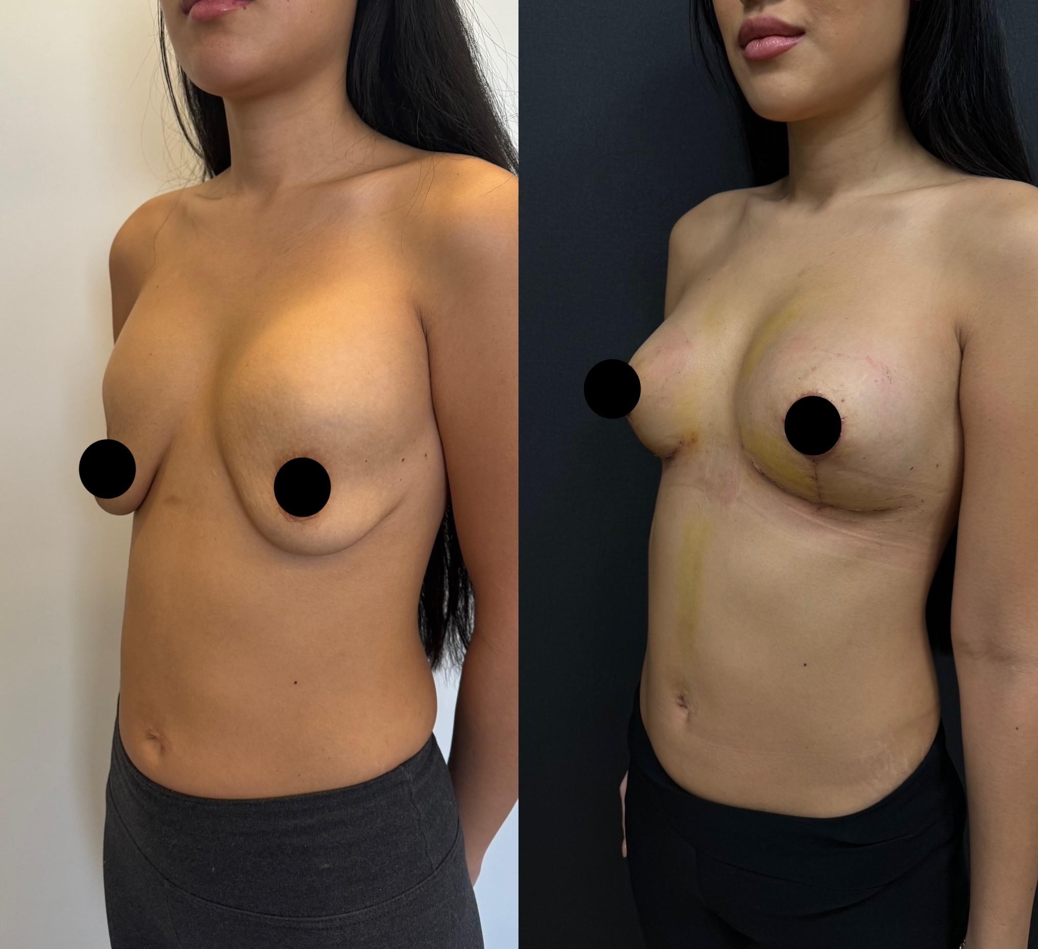
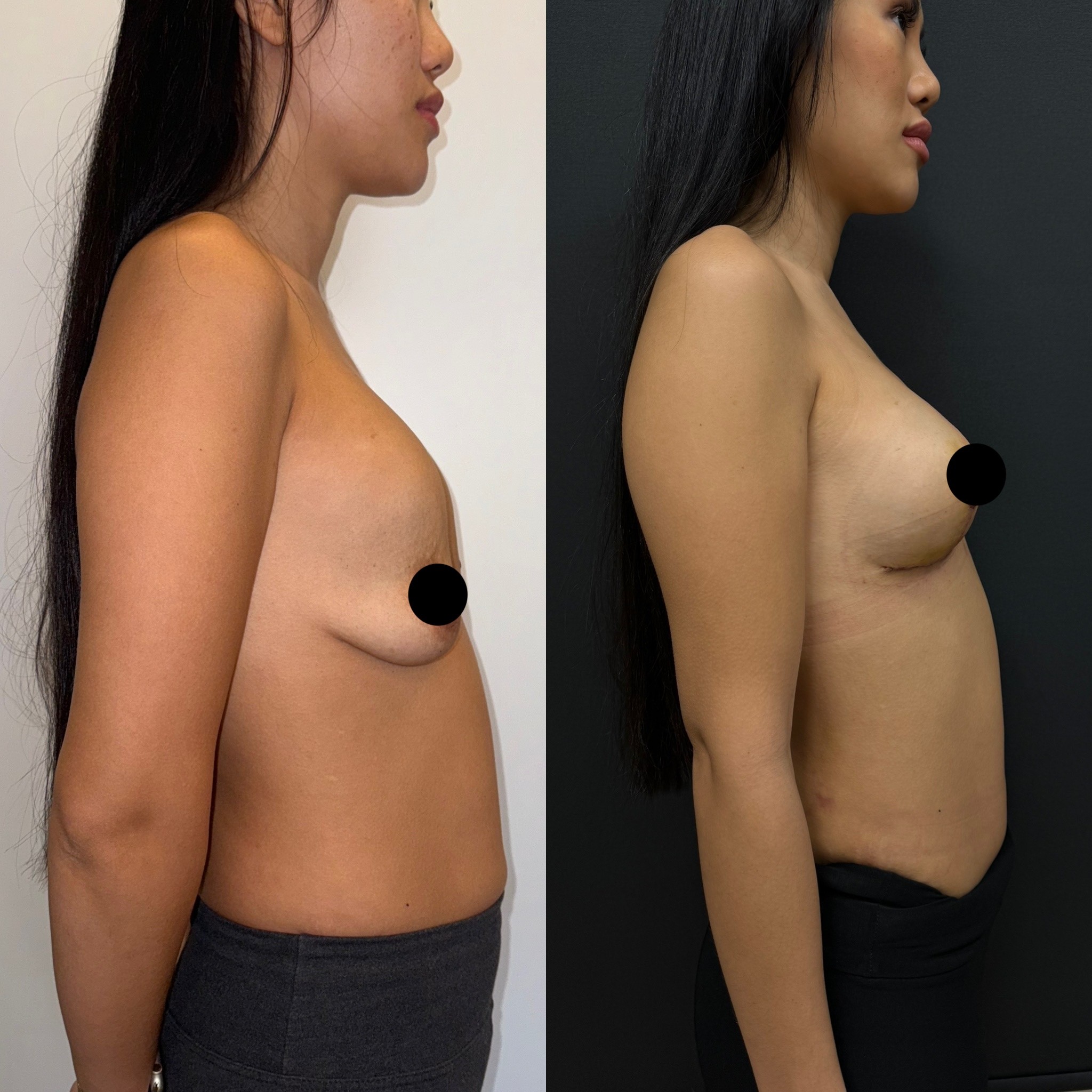
Waterfall deformity correction from full submuscular implants. Pocket conversion and muscle repair at 5 days by Dr. Cem Berkay Sinaci, Istanbul, Turkey.

Patient Overview
Patient: Ludmilla
Age: 37 years old
Gender: Female
Procedures: Implant removal from full submuscular pocket, pectoralis muscle repair and reattachment, creation of new prepectoral (over-the-muscle) pocket with new implants
Previous surgery: Breast augmentation with full submuscular implant placement (7 years prior, performed elsewhere)
After photos taken at: 5 days post-surgery
Case Description
Ludmilla's case represents the most severe form of waterfall deformity we have documented in our gallery — and it originated from a fundamental error in the original surgical technique. Seven years ago, she underwent breast augmentation with implants placed in a fully submuscular position — meaning the pectoralis major muscle was completely detached from its lower and lateral attachments and draped entirely over the implants. The result, over time, was a striking deformity: the implants sat high on the chest, completely encased behind the muscle, while her natural breast tissue descended below them under the influence of gravity. Each breast appeared to have two separate mounds — the implant above and the breast tissue below — creating the characteristic waterfall or double-bubble appearance.
This is distinct from Ayla's waterfall case, where partial submuscular placement allowed gradual tissue separation over time. Ludmilla's deformity was more pronounced because the implants were never in the correct position to begin with — full muscle coverage pushed them too high from the start, and seven years of gravity on the uncorrected tissue made the separation increasingly obvious.
Why Full Submuscular Coverage Creates Problems
In standard dual-plane breast augmentation, the pectoralis muscle covers the upper portion of the implant while the lower portion sits behind the breast tissue only. This partial coverage provides the benefits of muscle protection over the visible upper pole while allowing the lower pole of the implant to integrate with the breast tissue and move naturally.
Full submuscular coverage — where the muscle is released from its attachments and wrapped entirely around the implant — was a technique used more commonly in earlier decades of breast augmentation. The rationale was maximum implant concealment. The problem is that it creates a complete muscular barrier between the implant and the breast tissue, with two consequences that worsen over time.
First, the muscle holds the implant in an artificially high position. The pectoralis is attached to the clavicle and sternum superiorly — when it encases the implant completely, it tethers it to these upper anchor points. The implant cannot descend to the natural breast position because the muscle will not allow it.
Second, the breast tissue below the muscle is left completely unsupported by the implant. The implant's volume and projection — which should fill and support the breast mound — are trapped behind the muscular wall. The breast tissue in front of the muscle receives no structural benefit from the implant and continues to sag under gravity, just as it would in an unaugmented breast.
The result is exactly what Ludmilla experienced: two visually separate structures on each side of the chest. The high implant mound behind the muscle. The ptotic breast tissue in front of it. No integration between the two. A result that looked increasingly unnatural with each passing year.
The Correction: Complete Pocket Conversion and Muscle Repair
Correcting Ludmilla's deformity required undoing the original surgery entirely and rebuilding the breast from the ground up. This was the most extensive revision approach possible — not a modification of the existing pocket but a complete reconstruction of the implant-tissue relationship.
The first step was removing the implants from the submuscular pocket. Once the implants were out, the full extent of the muscle distortion was visible — the pectoralis had been detached from its inferior and lateral insertions to create the full-coverage pocket, leaving it in an abnormal, elevated position.
The second step was repairing the muscle. The pectoralis was reattached to its natural anatomical insertions, restoring it to its original position on the chest wall. This step is critical and is often overlooked in revision surgery. Simply closing the old pocket without repairing the muscle leaves the pectoralis in its displaced position, which can cause visible contour irregularities and animation deformity — where the muscle twitches or distorts the breast during chest movements.
The third step was closing the old submuscular pocket completely. The capsule that had formed around the implants over seven years was sutured shut, sealing the space to prevent any migration of the new implants back behind the muscle.
The fourth step was creating a new pocket in the prepectoral plane — directly in front of the repaired muscle and behind the breast tissue. The new implants were placed in this position, where they sit in the same compartment as the breast tissue and integrate with it as a single unit. There is no muscle barrier between the implant and the breast. They move together, age together, and present as one cohesive breast mound.
Why Prepectoral Rather Than Subfascial in This Case
In Ayla's waterfall correction, the new pocket was created in the subfascial plane — behind the fascial layer covering the muscle. In Ludmilla's case, the prepectoral plane was chosen instead. The distinction reflects the different surgical circumstances.
Ayla's pectoralis was intact and in its normal anatomical position — only a pocket conversion was needed, and the fascia provided a useful tissue layer for coverage. Ludmilla's pectoralis had been significantly detached and required formal repair and reattachment. After this repair, the fascial layer over the reconstructed muscle was not reliable enough to serve as a defined pocket boundary. The prepectoral plane — directly over the repaired muscle — provided a cleaner, more predictable space for implant placement.
At 37, Ludmilla has adequate breast tissue thickness to provide natural coverage over the implant in the prepectoral position. This is an important consideration — prepectoral placement requires enough overlying tissue to prevent visible implant edges and rippling. In patients with very thin tissue, the subfascial or even a new partial submuscular approach might be preferred.
Results at Day 5
At five days, the most important observation is that the double-mound deformity is gone. The implant and the breast tissue are now presenting as a single structure on each side. The two-separate-breasts appearance that defined the waterfall deformity has been replaced by a unified breast mound — swollen and elevated at this early stage, but cohesive.
The breasts are firm and edematous, as expected at day five of a revision procedure. The pectoralis repair adds an element of chest wall soreness that is not present in primary surgery or simpler revisions. The settling process ahead will be gradual — revision tissue, particularly tissue that has been through a major pocket conversion with muscle repair, takes longer to relax and conform to the new implant position.
Surgeon's Note
Ludmilla's case was the most technically involved breast revision I document in our gallery. The full submuscular placement she received seven years ago created a problem that could not be solved with a simple pocket adjustment — it required complete dismantling of the original surgical work and reconstruction from the muscle layer outward.
Repairing the pectoralis and restoring it to its anatomical position is a step that some revision surgeons skip, simply closing the old pocket and placing the new implant in front of it. I believe this is a mistake. Leaving the muscle in its displaced position creates a visible deficit on the chest wall and can produce animation artifacts. Taking the time to formally reattach the muscle restores the natural chest wall anatomy and provides a stable, well-defined surface for the new prepectoral implant to sit against.
At day five, the foundation is solid. The muscle is repaired, the old pocket is sealed, the new implants are in the correct plane, and the breast tissue and implant are functioning as a single unit for the first time in seven years. The settling process for this degree of revision will take three to six months, and I expect the final result to represent a dramatic improvement not only over the deformity but over the original augmentation result that Ludmilla had before the waterfall developed.
Frequently Asked Questions
What is the difference between full submuscular and dual-plane implant placement?
In dual-plane placement, the muscle covers only the upper portion of the implant while the lower portion sits directly behind the breast tissue. This allows the implant and breast tissue to move together naturally in the lower pole. In full submuscular placement, the muscle is wrapped entirely around the implant, creating a complete barrier between implant and breast tissue. This can cause the implant to sit too high and the breast tissue to descend separately, producing the waterfall deformity over time.
Can the pectoralis muscle be repaired after it was detached for implant placement?
Yes. The muscle can be reattached to its original anatomical insertions using sutures. This repair restores the normal chest wall contour and function. The muscle heals in its reattached position over several weeks and regains its normal strength. Formal muscle repair is an important step in revision surgery that prevents contour irregularities and animation issues.
Is prepectoral implant placement safe long-term?
Prepectoral (over-the-muscle) placement is increasingly used in both primary and revision breast surgery. Long-term studies show comparable complication rates to submuscular placement when patients are appropriately selected — specifically, when adequate tissue coverage exists over the implant. The advantages include elimination of animation deformity, more natural breast movement, and reduced postoperative pain compared to submuscular placement.
How long does recovery take after a complex revision like this?
The acute recovery — discomfort, swelling, activity restriction — follows a similar timeline to primary surgery, with most patients returning to normal daily activities within two to three weeks. However, the settling process may take longer than primary surgery because the tissue has been more extensively manipulated. Full settling and final result assessment typically occurs at four to six months rather than the three months typical of primary mastopexy-augmentation.