
Breast Augmentation for Glandular Ptosis at Day 3
Before and after breast augmentation correcting glandular ptosis without a lift at three days post-op. Early implant result by Dr. CBS in Istanbul, Turkey.
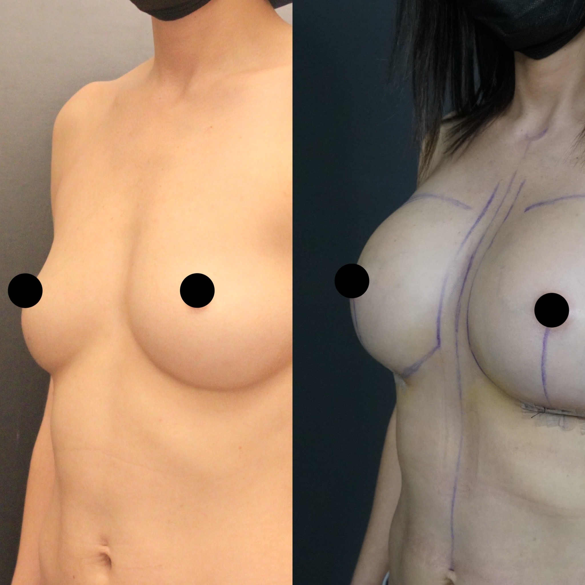


Patient Overview
Patient: Eida
Age: 36 years old
Gender: Female
Procedures: Breast augmentation with silicone implants (correcting glandular ptosis)
After photos taken at: 3 days post-surgery
Location: Istanbul, Turkey
Glandular Ptosis: The Sagging You Feel but Cannot Name
Many women sense that something about their breast shape has changed — the upper breast appears hollow, the volume seems to have shifted downward, and bras no longer fit the way they once did — yet their nipple still points forward at an apparently normal height. This paradox confuses patients who research breast sagging online and find that the clinical definitions focus almost entirely on nipple position relative to the inframammary fold. Their nipples sit where they should, so they conclude they do not have ptosis. They are wrong — they have glandular ptosis, and it is far more common than most women realise.
Eida, a thirty-six-year-old patient of Dr. Cem Berkay Sinaci, presented with this exact condition. Her nipple-areolar complex remained at an acceptable height on the chest wall, but the breast gland itself had descended within its skin envelope, leaving the upper pole deflated and the lower pole heavy. The breast had lost its youthful shape not because the skin had stretched catastrophically, but because the glandular tissue had migrated inferiorly under the influence of gravity, hormonal changes, and time.
Dr. Sinaci, a European board-certified plastic surgeon (FEBOPRAS) and active member of ISAPS and ASPS, identifies glandular ptosis through clinical assessment that goes beyond the standard ptosis grading systems. The distinction matters enormously because glandular ptosis, when correctly diagnosed, can often be corrected with augmentation alone — without the additional incisions and scars of a formal breast lift.
How Glandular Ptosis Differs from True Ptosis
Understanding the difference between glandular ptosis and true ptosis is essential for any woman considering breast surgery, because the distinction directly determines which operation she needs.
In true ptosis, the entire breast — gland, skin, and nipple — has descended. The skin envelope has stretched beyond its elastic capacity, and the nipple has migrated below the inframammary fold. Correcting this requires a mastopexy: the skin must be tightened, the nipple repositioned, and excess tissue removed. No implant alone can lift a nipple that has descended significantly.
In glandular ptosis, the skin envelope retains reasonable integrity. The nipple has not dropped below the fold. The problem is internal — the breast gland has slipped downward within an envelope that is still largely competent. When an implant is placed behind this descended gland, it pushes the tissue upward and forward, effectively redistributing the volume back into the upper pole and restoring the rounded shape that the breast has lost. The implant acts as an internal support structure, lifting the gland from behind without the need for external skin tightening.
Eida's anatomy at thirty-six placed her precisely in this category. Her skin quality remained adequate to support an implant, her nipple position was acceptable, and the primary deficit was volume displacement rather than envelope failure. Augmentation alone was the appropriate solution.
Why Day Three Photographs Are Clinically Valuable
Three days after breast augmentation is among the earliest time points at which before and after photographs are captured, and interpreting these images correctly requires understanding what the body is doing at this stage. Eida's day-three result is not a preview of her final outcome — it is a snapshot of her body's most acute healing response to surgery.
At seventy-two hours, the inflammatory cascade is approaching its peak intensity. Histamine and prostaglandins have increased capillary permeability throughout the operative field, allowing protein-rich fluid to accumulate in the interstitial spaces surrounding the implant. The breasts appear larger, firmer, and higher on the chest wall than they will at any subsequent point in recovery. The skin may appear taut and shiny from the underlying fluid pressure.
Despite these expected post-operative characteristics, the correction of Eida's glandular ptosis is already evident at day three. The upper pole, previously hollow and deflated, now shows volume. The breast mound sits higher and more forward on the chest wall. The overall breast shape has shifted from the bottom-heavy, descended appearance of glandular ptosis toward a more youthful, balanced contour. These improvements will become more refined as swelling resolves, but the fundamental correction is established the moment the implant is in position.
The Implant as an Internal Scaffold
The concept of using an implant to correct ptosis rather than simply add volume requires a shift in how patients think about breast augmentation. In Eida's case, the implant serves as a three-dimensional scaffold that occupies the space behind the descended gland and pushes it back into its ideal anatomical position.
This scaffolding effect depends on precise implant selection. The implant must have sufficient volume to fill the deflated upper pole and enough projection to push the gland forward into a natural-appearing contour. However, it must not be so large that it overwhelms the skin envelope's capacity to support it — an error that would create short-term fullness at the cost of accelerated stretching and eventual recurrence of the ptosis.
For Eida, Dr. Sinaci calculated the implant volume based on her tissue measurements and the degree of glandular descent observed during clinical examination. The goal was not maximum enlargement but optimal repositioning — choosing the volume that would restore her breast to a youthful shape while remaining within the structural limits of her existing tissue support.
The Recovery Arc from Day Three to Final Result
Patients who see their day-three result sometimes feel alarmed by the degree of swelling and the high implant position. Understanding the expected recovery timeline transforms this initial reaction from anxiety into informed patience.
Between days three and fourteen, the most noticeable change will be a progressive reduction in swelling. The breasts will begin to feel less tense, and the skin will lose its stretched, shiny appearance as interstitial fluid is reabsorbed through the lymphatic system. By two weeks, the breasts will look meaningfully different from what Eida sees now.
Between weeks two and six, the implants undergo their settling phase. The pectoralis muscle and surrounding soft tissue gradually relax their grip on the implant, allowing it to descend into its final position. The lower pole fills out, the upper pole softens, and the breast shape transitions from the early post-operative tightness to the soft, natural contour that characterises a well-healed augmentation.
By three months, the result is essentially final. The breast shape that Eida will live with has been established, the capsule around the implant has matured, and the glandular tissue has fully adapted to its new supported position. The correction of her glandular ptosis — which was already visible at day three — will by then be indistinguishable from a breast that developed with this shape naturally.
When Augmentation Alone Is Not Enough
Dr. Sinaci's decision to treat Eida with augmentation alone was based on careful clinical assessment that confirmed her glandular ptosis was within the range correctable by implant placement without mastopexy. This assessment, informed by his fellowship training with the internationally renowned plastic surgeon Raul Gonzalez in Brazil and advanced cadaver courses in Bangkok, draws on precise evaluation of nipple position, skin elasticity, gland volume, and the degree of descent.
Not every patient with glandular ptosis qualifies for this approach. When the nipple has begun to approach or cross the inframammary fold, when skin elasticity has diminished significantly, or when the degree of glandular descent is severe, augmentation alone will not produce an acceptable result. In these cases, a periareolar or vertical mastopexy combined with augmentation becomes necessary to reposition the nipple and tighten the envelope.
The ability to distinguish between patients who can be corrected with implants alone and those who require a combined approach is a clinical skill that protects patients from two opposite errors: undertreatment that leaves residual ptosis, and overtreatment that creates unnecessary scars on a breast that did not need them. For patients considering breast augmentation in Istanbul who have been told they have mild sagging, Eida's case demonstrates that a precisely planned augmentation may be all that is needed to restore the shape and fullness that glandular ptosis has taken away.